Mobile Menu
- Education
- Research
-
Students
- High School Outreach
- Undergraduate & Beyond: Community of Support
- Current Students
- Faculty & Staff
- Alumni
- News & Events
- Giving
- About

Researchers at the University of Toronto and Sinai Health have used a small worm to track how an animal’s brain changes throughout its lifetime, providing insight into how human brains may develop.
In research published today in the journal Nature, the scientists describe four basic patterns for the formation of new connections in the brain of the nematode worm Caenorhabditis elegans, or C. elegans.
“This is the first time that an entire brain’s structure is deduced and compared across developmental stages, from birth to adulthood,” said Mei Zhen, a professor of molecular genetics and physiology in the Temerty Faculty of Medicine at U of T and a senior investigator at the Lunenfeld-Tanenbaum Research Institute at Sinai Health. “These new findings have powerful implications for the fundamental rules that allow the brain’s developmental maturation to take place.”

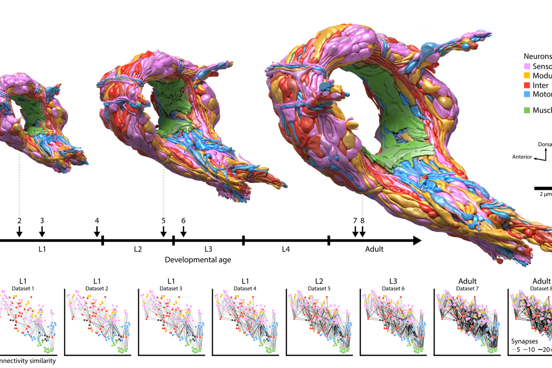
Zhen and her colleagues used state-of-art electron microscopes to reconstruct the full brain of eight C. elegans to learn how it changes with age. Although C. elegans is a relatively simple organism, many of the molecular signals controlling its development are also present in more complex organisms, including humans. It also has a life cycle of only two weeks, which makes it useful for scientists studying development.
“We examined every connection for every neuron and found many new connections are added as animals grow older,” said Zhen, who is also a professor of cell & systems biology in the Faculty of Arts & Science at U of T. “When we analyzed these connections like wires in a computational network, we discovered the new additions follow patterns and their collective changes serve one purpose — effective information processing.”
Daniel Witvliet, a former PhD student in molecular genetics in the Zhen lab, led the current study alongside scientists from Harvard University’s Center for Brain Science, including Professors Aravi Samuel and Jeff Lichtman.
The researchers looked at how the worm’s brain structure changed at single synapse resolution and at the geometry level as a whole, and discovered that a few structural properties in the newborn’s brain enable correct prediction of the wiring patterns in the mature brain.
“Our research shows that different parts of the brain have different degrees of flexibility,” Zhen said. “Knowing which part of the brain has more plasticity can help scientists come up with strategies to overcome genetic vulnerabilities to diseases during the brain’s development. We are humbled by and proud to continue the legacy from our scientific pioneers.”
The first wiring map of C. elegans was generated by John White and colleagues at MRC Cambridge more than 30 years ago, and it was a composite of partial maps from several different worms, including some at different developmental ages.
In addition to identifying some key patterns of the brain’s development, Zhen and her colleagues discovered substantial connectivity differences that make each brain unique.

They noticed consistent wiring changes between different neurons, where each change altered the strength of existing connections and in turn created new connections, ultimately altering how information was processed. Over the course of development, researchers found that central decision-making circuitry was maintained, while its sensory and motor pathways were substantially remodeled.
The research was funded by several organizations including the Canadian Institutes of Health Research, the U.S. National Institutes of Health and Human Frontier Science Program, along with support from Sinai Health Foundation and the Radcliffe Institute for Advanced Study at Harvard University.

