Mobile Menu
- Education
- Research
-
Students
- High School Outreach
- Undergraduate & Beyond: Community of Support
- Current Students
- Faculty & Staff
- Alumni
- News & Events
- Giving
- About

A new study from Sunnybrook Health Sciences Centre and the University of Toronto has shown that high-resolution ultrasound is as effective as MRI in detecting prostate cancer, potentially leading to faster diagnoses and fewer hospital visits, and freeing up the MRI for other uses.
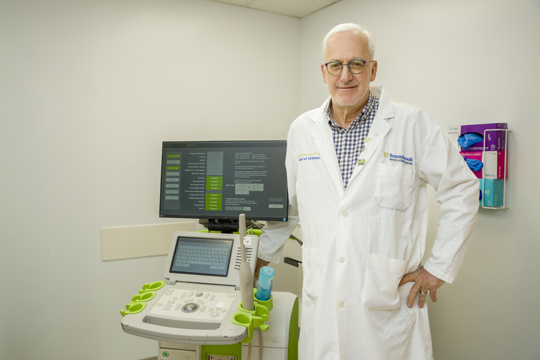
“This is practice changing for the diagnosis of prostate cancer. We can offer patients a one-stop shop, where they are imaged and then biopsied immediately, if required,” says Laurence Klotz, principal investigator of the trial, the Sunnybrook Chair of Prostate Cancer Research and a professor of surgery at U of T’s Temerty Faculty of Medicine.
“There’s no toxicity, no exclusions, it’s easier to use, and is much cheaper and more accessible; freeing up MRIs for hips and knees and all the other things they’re needed for.”
In a study published recently in JAMA, the researchers conducted the first randomized trial, named the OPTIMUM trial, to compared micro-ultrasound (microUS) with MRI-guided biopsy for prostate cancer.
The large international clinical trial involved 678 men who underwent biopsy at 19 hospitals across Canada, the US and Europe. Of these, half underwent MRI-guided biopsy, a third received microUS-guided biopsy followed by MRI-guided biopsy, and the remainder received microUS-guided biopsy alone.
In the study, microUS was able to identify prostate cancer as effectively as MRI-guided biopsy with very similar rates of detection across all three arms of the trial. There was little difference even in the group who received both types of biopsies, with the microUS detecting the majority of significant cancers.
Around a million prostate cancer biopsies are carried out each year in Europe, a similar number in the USA and around 100,000 in Canada. Most biopsies are conducted using MRI images fused onto conventional ultrasound, which enables urologists to target potential tumours directly, leading to more effective diagnosis.
MRI-guided biopsy is a two-step process (the MRI scan, followed by the ultrasound-guided biopsy) requiring multiple hospital visits and specialist radiological expertise to interpret the MRI images and fuse them onto the ultrasound.
MicroUS is a high-frequency ultrasound technology first pioneered in the 1990s by Stuart Foster, a senior scientist at Sunnybrook and professor of medical biophysics at Temerty Medicine. It uses higher frequency than conventional ultrasound, resulting in three times greater resolution images that can capture similar detail (of minute details within tissue) to MRI scans for targeted biopsies. Cheaper to buy and run compared to MRI, microUS could enable imaging and biopsy to be carried out during one appointment, even outside a hospital setting.
Clinicians such as urologists and radiologists can be easily trained to use the technique and interpret the images, especially if they have experience in conventional ultrasound.
Klotz notes that the results of the OPTIMUM trial could have a similar impact to the first introduction of MRI.
“When MRI first emerged and you could image prostate cancer accurately for the first time by doing targeted biopsies, that was a game changer,” he recalls.
“But MRI isn’t perfect. It’s expensive. It can be challenging to get access to it quickly and it requires a lot of experience to interpret properly. It also uses gadolinium which has some toxicity, and not all patients can have MRI, if they have replacement hips or pacemakers, for example. MicroUS is a Canadian innovation success story – the technology was developed right here at Sunnybrook.”
The trial was funded by Exact Imaging.

